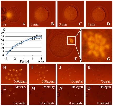
Figure 3. Staining of MII spindle using phycoerythrin and observation.
The antibody–phycoerythrin conjugate diffused immediately after microinjection into mouse oocytes (A) but the conjugate could bind to the MII chromosome array within a few minutes (B, C). Five minutes later, the chromosomes became sufficiently bright for enucleation (D). The brightness of MII chromosomes was measured by comparison with the cytoplasm; the luminance increased with time up to 8 min (E). Imaging could reveal individual chromosomes (F, G). Different concentrations of the phycoerythrin conjugate were microinjected into oocytes (H–K). The staining intensity was proportional to the concentration of conjugate used and 75 µg/mL of antibody was the minimum needed for clear observation. Fading of the phycoerythrin label was examined (L–O). (L, M) Images of phycoerythrin-injected oocytes observed using a conventional fluorescence microscope faded within 30 s. (M, N) When these samples were observed using the halogen light system, the image did not fade even when observed continuously for over 10 min.

