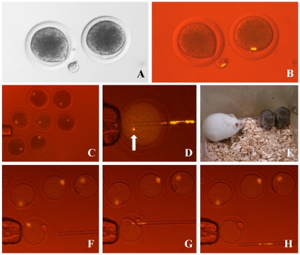
Figure 4. Enucleation of MII chromosomes from oocytes with fluorescence observation using halogen lamp.
The MII chromosomes of bovine oocytes are normally invisible by conventional microscopy because of dark lipid droplets in the ooplasm (A). After labeling with an H3S10ph antibody–phycoerythrin conjugate, the MII chromosomes could be recognized clearly and this allowed us to remove them along with minimal cytoplasm (B). MII chromosomes in porcine oocytes were detected by the same method (C). Some chromosomes (arrow) were left occasionally inside the cytoplasm of mouse oocytes after enucleation (D). Live and healthy cloned mice (brown) were obtained after enucleation using this method without any significant decrease in the success rate (E). Enucleation of the MII spindle from mouse oocytes with this system. (F) Before, (G) during and (H) after enucleation.

