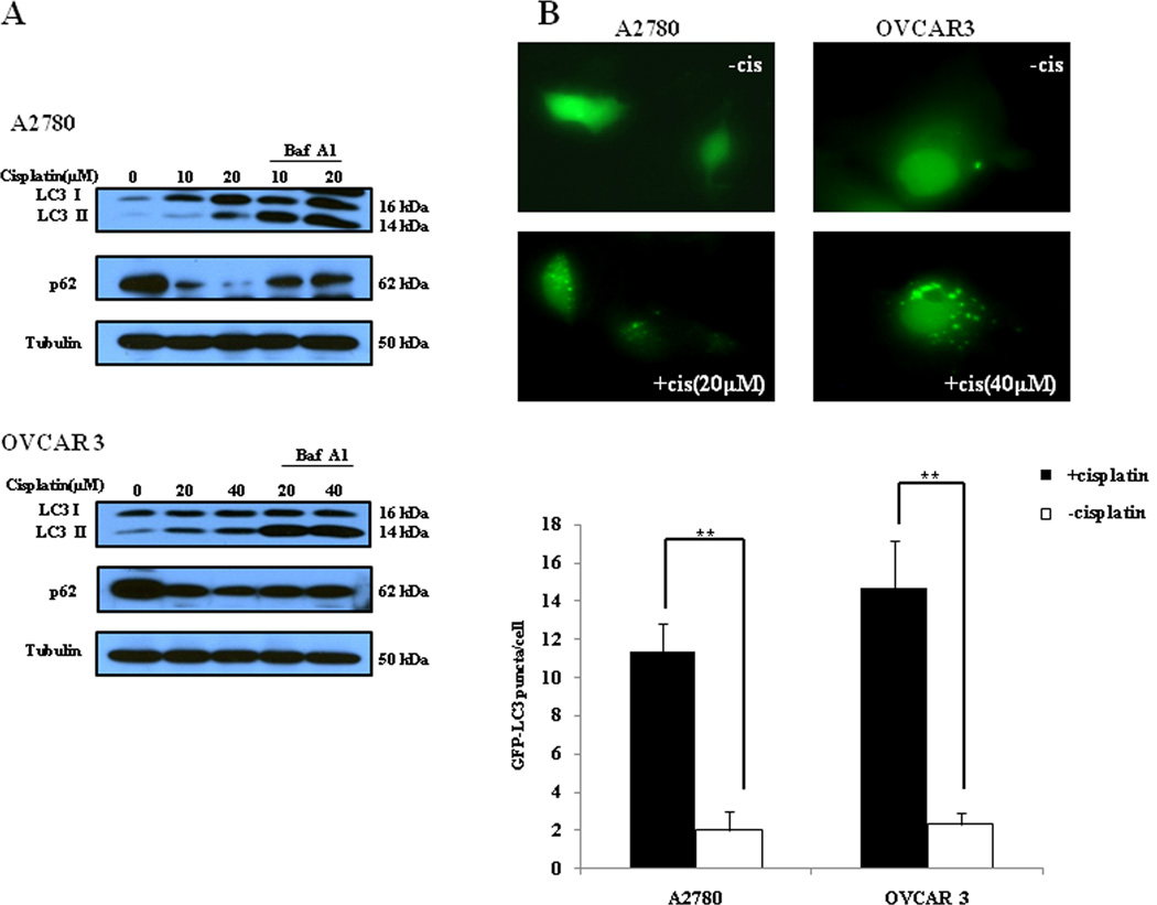
Figure 1. Cisplatin induces autophagy in ovarian cancer cells.
(A) A2780 and OVCAR3 cells were treated with the indicated concentrations of cisplatin for 24 h in the absence or presence of 10 nM of bafilomycin A1. At the end of treatment, cell lysates were prepared, resolved by SDS-PAGE, and subjected to Western blot analysis using anti-LC3, anti-p62 or anti-tubulin antibodies, respectively. Tubulin was used as a loading control. (B) A2780 and OVCAR3 cells were transfected with a GFP-LC3 plasmid, followed by treatment with the indicated concentrations of cisplatin for 24 h. At the end of treatment, the cells were inspected under a fluorescence microscope. Quantitation of the GFP-LC3 puncta was performed by counting 20 cells for each sample, and average numbers of puncta per cell were shown. The bars are the mean ± S.D. of triplicate determinations; results shown are the representative of three identical experiments. ** p < 0.01, t-test, cisplatin vs. vehicle.

