Figure 1.
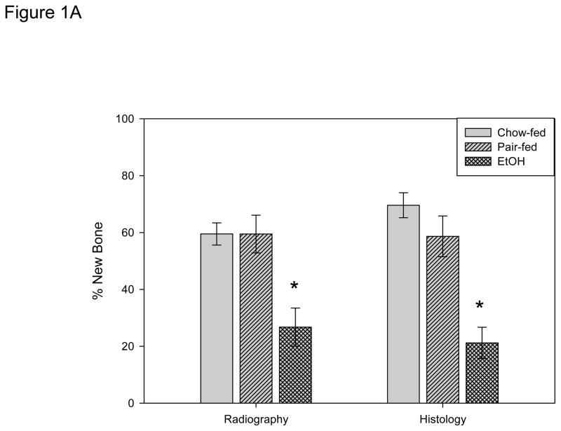
Figure 1A. New gap bone formation after DO in chow-fed, pair-fed and chronic EtOH fed B6 mice was assessed by single beam radiography and histology. Comparison of the distracted tibial radiographs demonstrated an EtOH–associated inhibition of % new bone mineralization (chow-fed: 59.5 ± 3.9% vs pair-fed: 59.5 ± 6.6% vs EtOH: 26.7 ± 6.7%, P= 0.004 & P<.001 respectively, F value=9.1 *). Further, analysis of histological sections supported the radiological analyses by revealing significant differences in new gap bone formation between both chow-fed (69.6 ± 3.4.4%) vs EtOH treated (21.2 ± 5.5%) mice, P< 0.001*, and pair-fed (58.6 ± 7.2%) vs EtOH treated mice, P<0.001, F value=17.4 *.
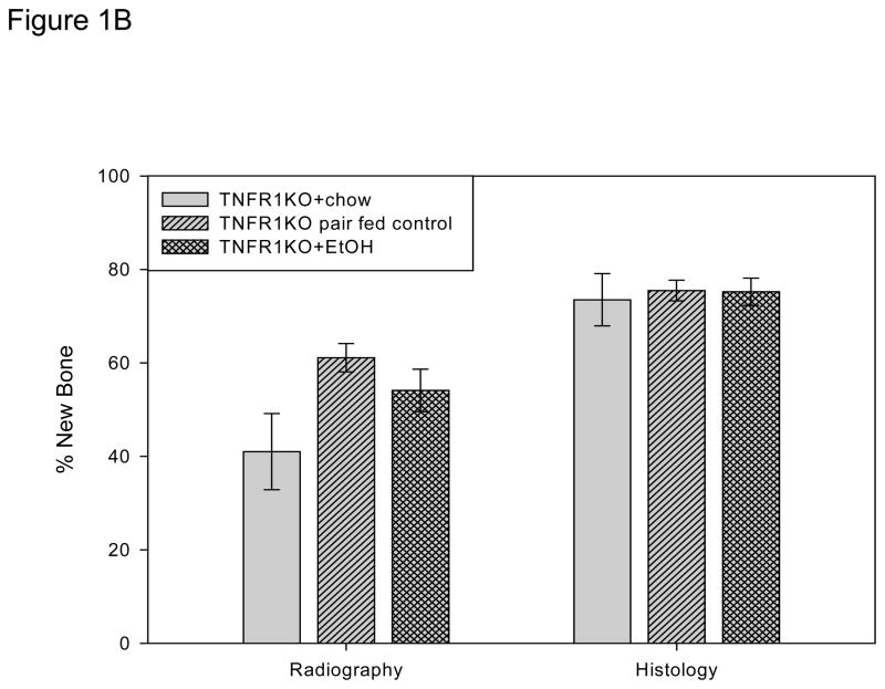
Figure 1B. New gap bone formation after DO in chow-fed, pair-fed, and chronic EtOH fed TNFR1 KO mice was assessed by single beam radiography and histology using One-Way ANOVA. Comparison of the distracted tibial radiographs demonstrated a trend for lower values in the chow fed control which reached significance vs the pair-fed (P<0.05) but not vs the EtOH treated mice (P=0.09). The EtOH vs the pair-fed values were not significant (P=.3). The values for the three groups were chow-fed: 41.0 ± 8.2% vs pair-fed: 61.1 ± 3.0% vs EtOH: 54.1 ± 6.7%. However, analysis of histological sections demonstrated no significant differences between the groups (chow-fed (73.5 ± 5.6%) vs pair-fed (75.5 ± 2.2%) vs EtOH treated: 75.2 ± 2.9%) (P=0.916, F value=.09).


