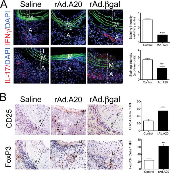
Figure 4.
Overexpression of A20 favors an immunoregulatory T Cell response. A. Representative immunofluorescence staining shows decreased adventitial expression of IFNγ and IL-17 in rAd.A20-treated aortic allografts, as compared to saline and rAd.βgal-treated vascular allografts. Color codes are as follows: red-IFNγ or IL-17, blue-DAPI nuclear staining, green-elastic laminae auto-fluorescence. Graphs depict grading scores for the IF staining within vascular allograft. Each bar represents the mean±SE IF score from 3 mice in the rAd.A20-treated group and 4 mice in the control group (2 saline and 2 rAd.βgal-treated) for IFNγ statining, and from 4 mice in the rAd.A20-treated group and 6 mice in the control group (3 saline and 3rAd.βgal-treated) for IL-17 staining. B. Representative immunohistochemistry photomicrographs show increased number of CD25+ and FoxP3+ T cells/HPF in rAd.A20-treated aortic allografts, as compared to controls (saline and rAd.βgal treated) vascular allografts. Each bar represents the mean±SE number of CD25+ and FoxP3+ cells/HPF from 4 mice in the rAd.A20-treated group and 5 mice in the control group (2 saline and 3 rAd.βgal-treated.) I-intima, M-media and A-adventitia, original magnification X400. *p<0.05, **p<0.01, ***p<0.001.

