
Figure 5.
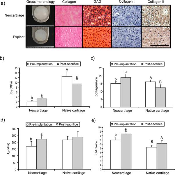
Properties of constructs post-sacrifice after in vivo culture in nude mice. Self-assembled articular cartilage was cultured for 4 weeks with TGF-β1 treatment during weeks 1&3 and chondroitinase-ABC (C-ABC) administration at day 14. Neocartilage was then implanted in nude mice for 4 weeks with condylar cartilage explants as controls (n=4). (a) Gross morphology and histology using Picrosirius Red for collagen, Safranin-O/fast green for glycosaminoglycans, and immunohistochemistry for collagen I and collagen II. Scale bar is 200 μm. (b) Post-sacrifice tensile stiffness increased for neocartilage while explant stiffness decreased. (c) Collagen content increased for constructs post-sacrifice while it decreased for native cartilage. (d) Compressive stiffness increased for constructs but did not significantly change for explants. (e) Glycosaminoglycan (GAG) content increased for both native tissue and neocartilage post-sacrifice. Bars labeled with different letters exhibit significant differences (p < 0.05).