
Figure 4.
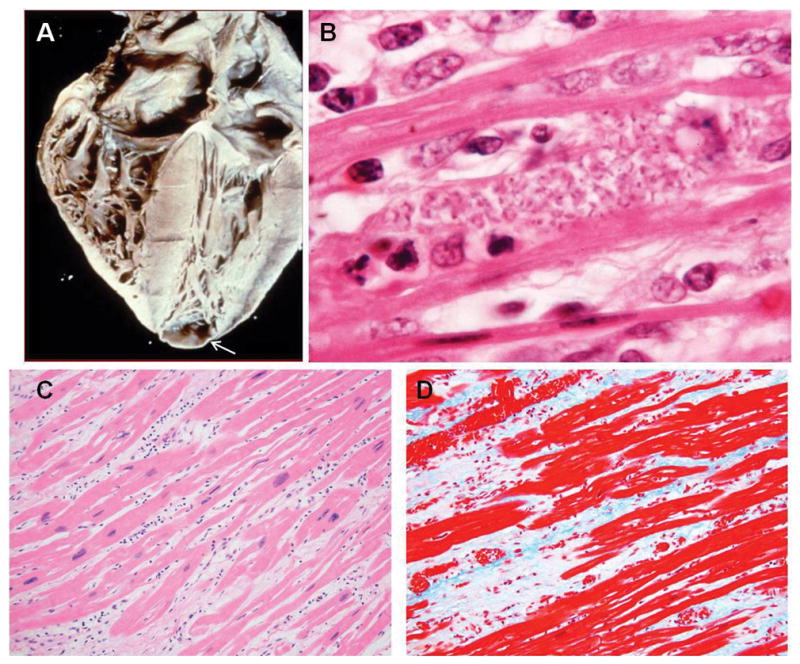
A: Heart of a patient with chronic Chagasic cardiomyopathy. There is four-chamber enlargement of the heart. Note the apical aneurysm (arrow). B: Rare pseudocyst in the myocardium of a patient with chronic Chagasic cardiomyopathy. C: H&E stained myocardium of a patient with chronic Chagasic cardiomyopathy showing bands of fibrous tissue. D: Myocardium of the same patient stained with Trichrome showing massive fibrosis. 4B from the collection of Herman Zaiman’s “A Presentation of Pictorial Parasites”, with permission from the American Society of Tropical Medicine and Hygiene.