
Figure 6.
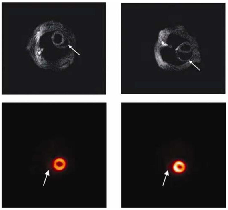
Cardiac imaging of Trypanosoma cruzi infected mice: A: Cardiac MRI of an uninfected mouse. The right ventricle is normal in size (arrow). B: Cardiac MRI of a Trypanosoma cruzi-infected mouse 100 days post infection. Note the enlarged right ventricle (arrow). C: Cardiac microPET of an uninfected mouse showing the right ventricle (arrow). D: Cardiac microPET showing an enlarged right ventricle (arrow) in heart of a mouse 60 days post infection. From Prado et al (Ref. 139) with permission.