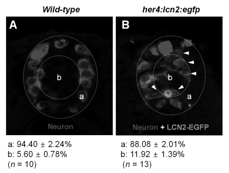
Figure 4.
The expression of LCN2 attracts developing neurons toward medial position of the spinal cord in zebrafish. The wild-type embryo (A) or her4:lcn2:egfp-injected transgenic embryo (B) was labeled with an anti-Hu antibody to detect neurons at 24 hpf. Arrowheads indicate neurons near the lcn2:egfp-expressing cells. Dotted lines indicate a lateral margin (a) and medial position (b) of the spinal cord. Numbers indicate percentage of neuronal cells in each region. All images are transverse sections of zebrafish spinal cord, dorsal to top. The quantification of cell migration was done by enumerating the migrated cells as described in the Materials and Methods section. The results are mean±SD.

