Figure 1.
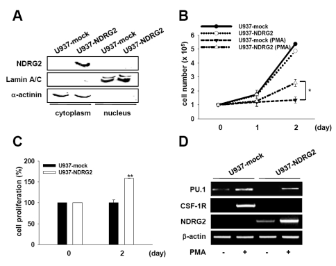
Overexpression of NDRG2 in U937 cells. (A) Cell lysates were fractionated into cytoplasmic and nuclear components. Western blotting using an anti-NDRG2 antibody was performed for each fraction. Lamin A/C and α-actinin were used as control for protein loading as well as marker proteins for nuclear and cytoplasmic fraction, respectively. (B, C) U937-mock and U937-NDRG2 cells were treated with 0.1 µg/ml PMA for 48 h. Cell viability was determined using either the trypan blue exclusion assay (B) or the CCK-8 assay (C). The results represent the means±SD of duplicates. *p<0.05, **p<0.01. (D) PU.1, CSF-1R, and NDRG2 mRNA levels were measured by RT-PCR. β-actin was used as a loading control.

