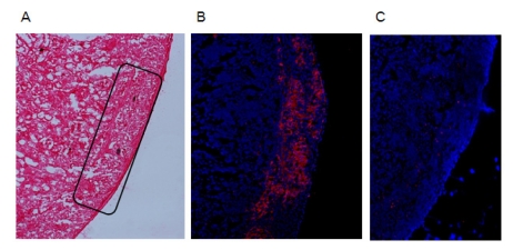
Figure 3.
Histological analysis of islet graft. Removed kidney containing grafted islet of recipient in Fig. 3B was frozen-sectioned as 5µm thickness and acetone-fixated. (A) H&E staining was done to the section. Rectangular area indicates islet region. (B) Insulin staining was done with primary guinea pig anti-insulin antibody and secondary Alexa Fluor 555-conjugated anti-guinea pig IgG antibody, subsequently. Red spots indicate plenty of stained insulin in islet region. Blue spots are nuclei of cells resulted by DAPI staining. (C) As a control of '(B)', secondary antibody staining without primary antibody did not result in non-specific red spots. Magnifications are ×100.

