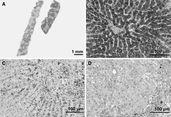
Fig. 2.
Histological appearance of copper-associated hepatitis in different dog breeds. Slides are stained with rubeanic acid and hematoxylin counterstain. a Liver biopsy of a female Bedlington terrier clearly showing centrolobular distribution of copper. b Liver biopsy of a female Bedlington terrier, 3 years of age with a liver copper value of 11,500 μg/g dwl copper. Massive amounts of copper granules are visible mainly in hepatocytes but also in Kupffer cells. The central vein is located in the middle of the picture. c Liver biopsy of a female Labrador, 5 years of age with a liver copper concentration of 2,360 μg/g dwl. Copper granules are present in hepatocytes and macrophages in the centrolobular area. The centrolobular region is characterized by loss of hepatocytes, mild fibrosis, and moderate numbers of lymphocytes and plasma cells. d Liver biopsy of a female Dobermann, 6 years of age with a liver copper value of 1,700 μg/g dwl. The centrolobular area (bottom right of the picture) is characterized by mild fibrosis with multifocal accumulation of macrophages containing lipofuscin pigment and copper granules. Furthermore, this area shows moderate infiltration with lymphocytes. Hepatocytes in the centrolobular region contain moderate amounts of copper granules

