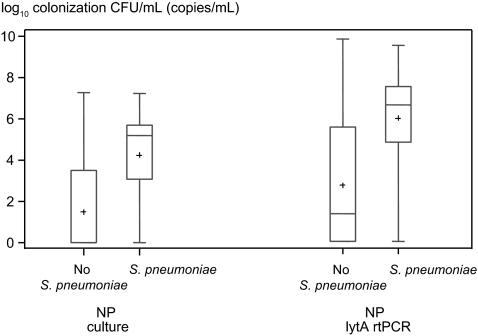
Figure 1.
Quantitative colonization densities in human immunodeficiency virus–infected patients with community-acquired pneumonia. Pneumococcal colonization densities are shown as colony counts, obtained with nasopharyngeal (NP) swab cultures and lytA real-time polymerase chain reaction (rtPCR) of NP swab samples. Counts are depicted after logarithmic transformation for those who had Streptococcus pneumoniae identified by the composite diagnostic standard and those who did not. Plus signs represent means; lengths of boxes, interquartile ranges between 25th and 75th percentiles; horizontal lines in boxes, medians; and whiskers, minimum and maximum values (t test, P < .001 for NP counts from cultures and for lytA rtPCR; Wilcoxon rank sum test [used after assignment of value 1 to the colony count for those who were not colonized], P < .001 for NP counts and lytA rtPCR). Abbreviations: CFU, colony-forming units; NP, nasopharyngeal; rtPCR, real-time polymerase chain reaction.

