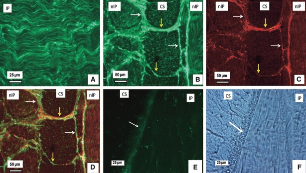
Fig. 3.

Dual immunostained LTBP-2 and fibrillin-1 in the outer annulus fibrosus. (A) Parallel LTBP-2 fibres (green) in the inplane (IP) lamella. (B–D) Typical dual immunostaining of a section with (B) showing LTBP-2 (green), (C) fibrillin-1(red) and (D) the merged image of (D) and (E). LTBP-2 appears highly co-localised with fibrillin-1; together they are densely organised in the interlamellae spaces (white arrows) and also at the boundaries of collagen bundles (yellow arrows). (E–F) Hyaluronidase overnight pre-digestion removed LTBP-2 (green in E), but not collagen structure (F), with white arrows indicating interlamellae. CS, cross-sectioned lamella; nCS, nearly cross-sectioned lamella; IP, inplane lamella; nIP, nearly inplane lamella.
