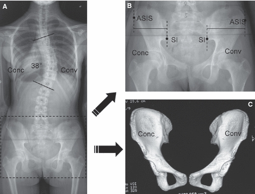
Fig. 1.

(A) The PA radiograph of a 14-year-old female patient with AIS with a main thoracic curve of 38°. (B) The location of the hipbone landmarks. The inferior ilium at the sacroiliac joint (SI) and anterior superior iliac spine (ASIS) are indicated. The distance between two plumb lines through the SI and ASIS was defined as the SI–ASIS. The concave/convex ratio of the SI–ASIS in this patient was 0.87. (C) The 3D reconstruction of the hipbones after the CT scan. The hipbone volume was 215.2 cm3 at the concave side and 215.7 cm3 at the convex side.
