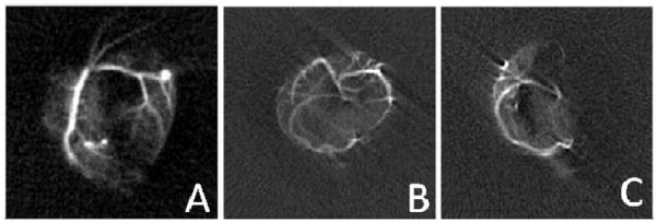
Figure 1.
MR images acquired using microbubbles with 3He gas cores as contrast agents. A) Image acquired after introduction of microbubbles into the left coronary artery. B) A transverse image projection acquired after microbubbles injected in both coronary arteries prior to the occlusion of the left coronary artery. C) A transverse image projection acquired after microbubbles injected in both coronary arteries after the occlusion of the left coronary artery. The field of view in these three images is approximately 14 cm. Adapted from 32. (Reprinted from Acad. Radiol., 9, (Suppl. 2), V. Callot, E. Canet, J. Brochot, H. Humblot, A. Briguet, H. Tournier and Y. Cremillieux, ‘Hyperpolarized helium3 encapsulated in microbubbles: a new class of blood pool MRI contrast agent’, S501–S503, 2002, with permission from Elsevier.)

