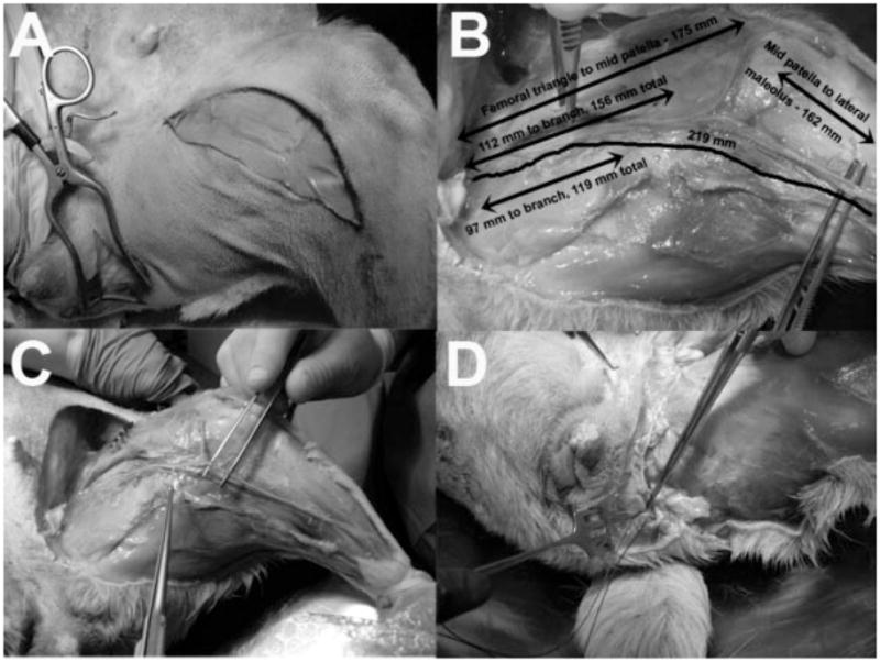
Fig. 1.

Feasibility study in cadaver dogs showing the dissection technique. This was performed prior to surgical procedures. A: Surgical incision sites on anterior thigh for access to the anterior femoral nerve branch and femoral nerve motor branches to the sartorius muscle and articularis genu muscle which is the nervus saphenous pars muscularis in canines,8 and to the lateral perineum for access to the pudendal nerve in Alcock’s canal. B: Various relevant dimensions of the femoral motor nerve branches. C: Mobilization of the femoral motor nerve is displayed. D: The femoral motor nerve reaches the pudendal nerve as it emerges from Alcock’s canal.
