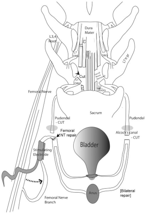
Fig. 2.
Diagram of the surgical transfer and repair methology. A spinal leminectomy was performed. Sacral ventral roots (S1, 2) were selected by their ability to stimulate sphincter contraction and transected. On the right side of the diagram, Alcock’s canal (gray oval) is shown. This is the site at which the pudendal nerve passes superficially into the perineum. On the left side of the diagram, the origin of nervus saphenous pars muscularis as part of the femoral nerve branches is shown. Bilaterally, motor branches of posterior branches of the femoral nerve were mobilized, transferred (dotted arrow) and attached by end to end anastomosis to the distal end of the transected pudendal nerves as they emerged from the pudendal canal in the perineum (as shown on the left), and enclosed in unipolar nerve cuff electrodes with leads to RF micro-stimulators. The external urethral spincter is shown at the base of the bladder, and the external anal sphincter is represented by the oval labeled as anus. The spinal cord laminectomy and root transections are the same as previously described and diagramed in Ruggieri et al.5. Abbreviatons: CG, coccygeal roots; DRG, dorsal root ganglion; L, lumbar toots; S, sacral roots; tr pr, transverse processes.

