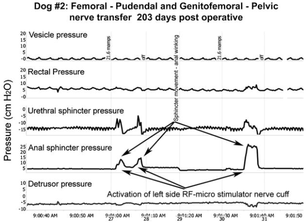
Fig. 3.
Functional electrical stimulation. Representative urodynamic pressure recordings during FES (arrows) via the implanted RF micro-stimulator with a lead to a unipolar nerve cuff electrode surrounding the transferred femoral motor nerve branches 1 cm proximal to the anastomosis with the pudendal nerve branches on the left side in dog #2. Foley balloon catheters (12 Fr, 5 ml balloon) were passed through the vesicostomy for bladder filling and through the urethra for vesicle pressure measurement using external transducers. Similar pressure transducers connected to the lumen of 10 Fr ballon catheters were passed into the rectum and into the urethral and anal sphincters for monitoring rectal, urethral sphincter and anal sphincter pressures. The bottom detrusor pressure trace is derived by subtracting the rectal pressure (as a surrogate for abdominal pressure) from the top trace for intravesical pressure. Similar increases in urethral and anal sphincter pressures during stimulation were observed for 5 of the 6 transferred femoral motor nerves.

