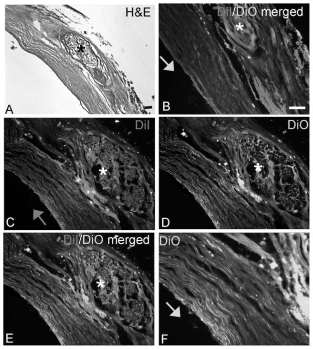
Fig. 5.
DiI and DiO labeling confirmed axonal regrowth across the nerve repair site. A: H&E stained section showing a repaired nerve at the site of reanastomosis, as indicated by the suture (*). B: DiO (green axonal dye) was placed proximal to site of reanastomosis while DiI (red axonal dye) was placed distal to the site of reanastomosis, as indicated by the arrow. C: Higher power micrograph showing DiI-labeled axons crossing the repair site. D: Higher power micrograph showing Di-labeled axons crossing the repair site. E: Merged (C) and (D). F: Even higher power micrograph showing DiO-labeled axons crossing the repair site. Scale bar = 50 μm.

