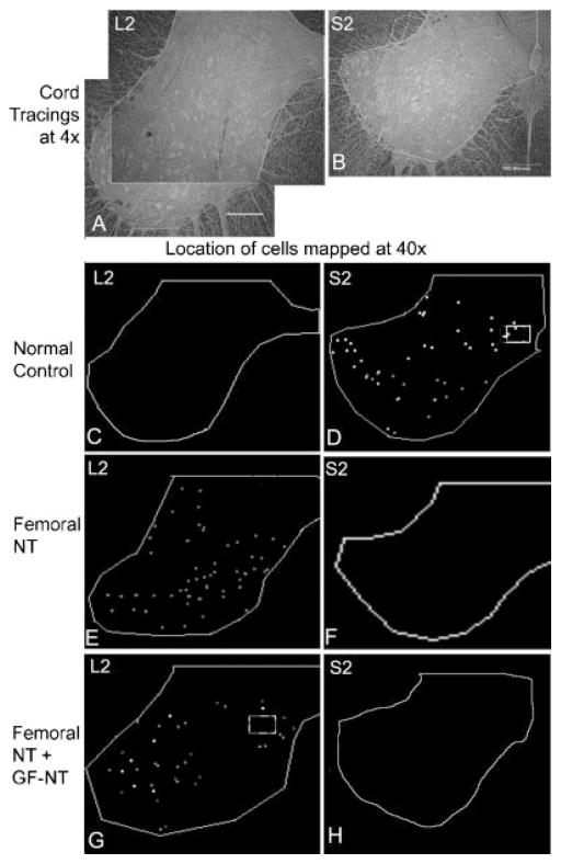
Fig. 6.
Location of neuronal cell bodies in the spinal cord retrogradely labeled with fast blue, fluororuby, or fluorogold after injection into the urethral sphincter, anal sphincter, or urinary bladder, respectively. A: Micrograph of an unstained bright-field section (taken with a ×4 objective) of a lumbar 2 (L2) spinal cord segment showing tracing around area in which neurons were counted. In this spinal cord segment, two larger areas were counted, as indicated, and the data combined. B: Micrograph of a sacral 2 (S2) spinal cord segment showing tracing around area analyzed (4.5 mm2). Scale bars in (A) and (B) = 500 μm. C, D: Representative tracings showing the location of fluororuby (red), fast blue (blue), and fluorogold (green after specific detection with Ki-67 antibody-W (Fluorochrome, LLC) and Cy2 secondary antibody) retrogradely labeled cell bodies in an S2 segment in a normal control. No retrogradely labeled neurons were detected in lumbar segments of normal controls. E, F: Representative tracings showing the location of retrogradely labeled cells in L2 and S2 segments in an animal in which nervus saphenous pars muscularis (L2–4) branches of the femoral nerve were transferred to the pudendal nerve (femoral-NT). No retrogradely labeled neurons were detected in sacral segments of Femoral-NT. G, H: Representative tracings showing the location of retrogradely labeled cells in L2 and S2 segments in an animal in which femoral nerve branches were transferred from the thigh to the pudendal nerve in Alcock’s canal (femoral-NT), and in which genitofemoral nerve branches were transferred intra-abdominally to the pelvic nerve branch to the bladder (GF-NT).

