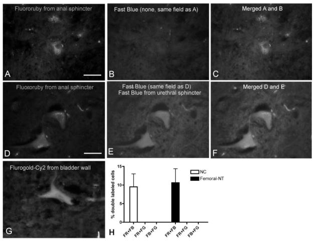
Fig. 8.
Examples of neuronal cell bodies in the spinal cord retrogradely labeled with fluororuby (A, D), fast blue (B, E), and fluorogold-Cy2 (G) after injection into the external urethral sphincter, anal sphincter or urinary bladder, respectively. Fluororuby-labeled neurons shown in (A) are not double-labeled for fast blue (B) in the merged image (C), while fluororuby-labeled neurons shown in (D) are double-labeled with fast blue (E) in the merged image (F). The graph in panel (H) shows that the percent of double-labeled neurons in sacral segments of normal controls (NC) and in the lumbar segments of femoral to pudendal nerve transfer cords was similar. Scale bar = 50 μm.

