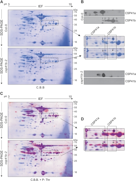
Fig. 4.
CSP41 proteins are post-translationally modified. (A) Stromal proteins (500 μg) from WT (Col-0) and csp41b-3 plants were fractionated by IEF in the first dimension and by SDS–PAGE in the second. Proteins were transferred to PVDF membranes and stained with Coomassie brilliant blue (C.B.B.). (B) Sections of A containing CSP41 proteins stained with C.B.B. or immunodecorated with specific antibodies raised against CSP41a or CSP41b are shown. Arrows indicate the main CSP41a and CSP41b protein species. (C) The membranes from A were immunodecorated with antibodies against phosphorylated threonine residues; the detected signals were converted to red and merged with the image shown in A. Phosphorylated proteins are shown in purple, and highly phosphorylated proteins in red. (D) Sections of C containing CSP41 proteins. The asterisk marks glyceraldehyde-3-phosphate dehydrogenase B (Goulas et al., 2006). This protein, together with RbcL and RbcS, has been previously found to be phosphorylated (Lohrig et al., 2009; Reiland et al., 2009). Arrows indicate the main CSP41a and CSP41b protein species. (A–D) At least two independent experiments were performed with similar results.

