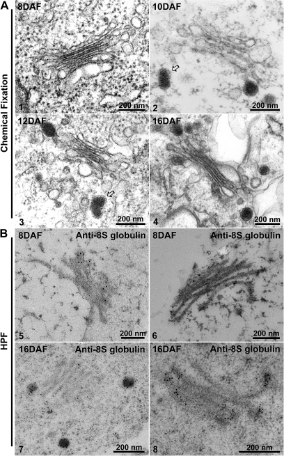
Fig. 7.
Ultrastructural and IEM analysis of Golgi and dense vesicles (DVs) in developing mung bean seeds. Structural EM analysis. Developing mung bean cotyledons at various stages (8–16 DAF) as indicated were collected, followed by chemical fixation for structural EM analysis. (Panels 1–4) The arrows in panels 2 and 3 point to clathrin caps at the DV (see the Results). IEM analysis. Developing mung bean cotyledons at various stages (8 DAF and 16 DAF) as indicated were collected, followed by high-pressure frozen/freeze-substitution (HPF) and IEM labelling using 8S globulin antibodies as indicated. Scale bars=200 nm.

