Fig. 4.
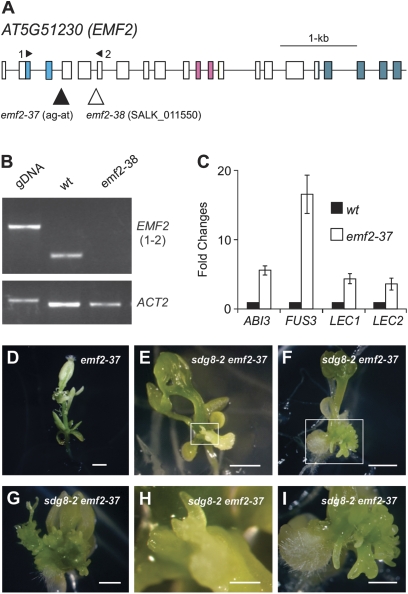
Phenotypes of the sdg8-2 emf2-37 double mutants. (A) Structure of the EMF2 gene and the location of mutation/T-DNA insertion sites of emf2 alleles. Boxes and lines represent exons and introns, respectively. The shaded boxes represent the conserved protein domains (from left to right): conserved N-terminal basic domain, C2H2-type zinc finger domain, and C-terminal acidic-W/M domain. The mutation in emf2-37 is ‘G’ to ‘T’ at base pair 20 8247 27 on chromosome 5. (B) RT-PCR analysis of the expression of EMF2 in the wild type and emf2-38 mutants. The primers used are indicated in (A). Genomic DNA (gDNA) was included as a size control for RT-PCR products, and Actin2 was used as an internal control. (C) qRT-PCR analysis of ABI3, FUS3, LEC1, and LEC2 genes in seedlings (aerial portion) of emf2-37 mutants grown for 15 d on MS agar. Wild-type (Col) RNA levels are designed as 1-fold. The expression of Actin-8 was used as an internal control. The mean and standard error were determined from three biological replicates, each of which was conducted in triplicate. (D–I) Morphological phenotypes of emf2-37 single (D) and sdg8-2 emf2-37 double mutants at different growth phases on MS agar (E, 16 d; F, 25 d; G, 32 d). (H) and (I) are close-up images of the boxed areas in (E) and (F), respectively. Bar=1 mm. (This figure is available in colour at JXB online.)

