Fig. 6.
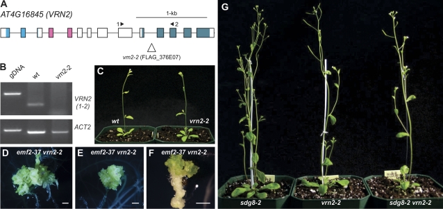
Characterization of a new vrn2 allele and phenotype of the sdg8 vrn2 double mutants. (A) Structure of the VRN2 gene and the location of the T-DNA insertion site of the vrn2-2 allele. Boxes and lines represent exons and introns, respectively. The shaded boxes represent the conserved protein domains (from left to right): the conserved N-terminal basic domain, the C2H2-type zinc finger domain, and the C-terminal acidic-W/M domain. (B) RT-PCR analysis of the expression of VRN2 in the wild type and the vrn2-2 mutant. The primers used are indicated in (A). Genomic DNA (gDNA) was included as a size control for RT-PCR products, and Actin2 was used as an internal control. (C) Phenotype comparison of the vrn2-2 mutant at 25 d with the wild type (Ws ecotype). (D–F) Morphological phenotypes of the emf2-37 vrn2-2 double mutants grown on MS agar (D and E, 30 d; F, 20 d). Bar=1 mm. (G) Phenotype comparison of the sdg8-2 vrn2-2 double mutant with the sdg8-2 and vrn2-2 single mutants at 30 d. (This figure is available in colour at JXB online.)

