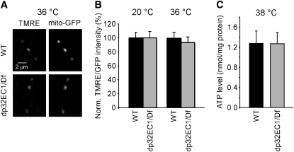
Figure 8 .
dp32EC1 mutant retains wild-type mitochondrial membrane potential (ΔΨm) and ATP production. (A, B) ΔΨm measurements using tetramethylrhodamine ethyl ester perchlorate (TMRE) and GFP targeted to the mitochondrial matrix (mito-GFP). (A) Examples of live confocal images of TMRE at DLM neuromuscular synapses exhibiting neuronal expression of UAS-mito-GFP in a WT or dp32EC1 genetic background. Mito-GFP serves as a mitochondrial marker within presynaptic terminals. (B) dp32EC1 retains wild-type ΔΨm. The ratio of TMRE to GFP (TMRE/GFP) from the same region of interest was used to represent the relative ΔΨm, which should be independent of mitochondrial size, shape, and orientation. The relative ΔΨm from dp32EC1 is shown as a mean percentage of that from WT. The ΔΨm in dp32EC1 is not significantly different from WT at either temperature. The respective ratios of TMRE to GFP at WT and dp32EC1 synapses were 1.32 ± 0.11 (n = 26) and 1.30 ± 0.11 (n = 23) at 20°. The corresponding values at 36° were 0.60 ± 0.06 (n = 22) and 0.56 ± 0.05 (n = 24). (C) Head homogenates from dp32EC1 contain wild-type levels of ATP. Head homogenates prepared from WT and dp32EC1 flies were exposed to 38° for 10 min and subjected to a luciferin-luciferase ATP detection assay. The relative ATP levels were calculated by dividing the luminescence by the total protein concentration. The relative ATP levels in WT and dp32EC1 were 1.28 ± 0.24 nmol/mg protein (n = 5) and 1.27 ± 0.23 nmol/mg protein (n = 5), respectively, and not significantly different.

