Fig. 2.
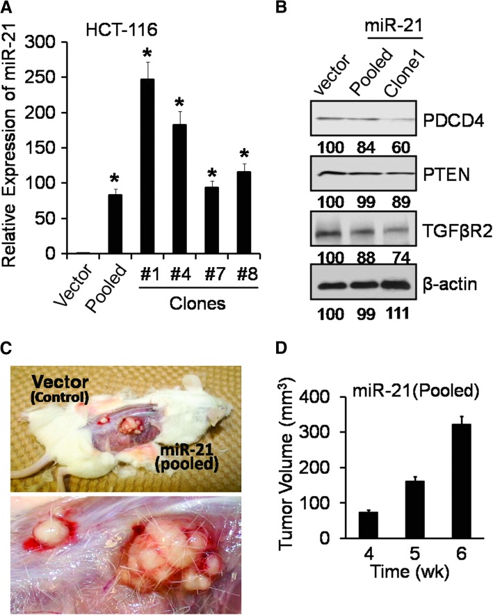
Overexpression of human miR-21 in colon cancer HCT-116 cells increases tumorigenic potential in SCID mice (A) qRT–PCR showing upregulation of mature miR-21 in HCT-116 clones, derived from the cells stably transfected with pCMV-miR-21 plasmid or the corresponding vector (controls), *P < 0.001. (B) Western blot showing downregulation of TGFβR2, PDCD4 and PTEN by miR-21. β-Actin was used as loading controls. The numbers represent percent of corresponding control normalized to β-actin. (C) Representative photographs showing the anatomical intact xenograft tumor in female SCID mice as observed at the end of 6 weeks of tumor growth after injecting HCT-116 cells stably transfected with pCMV-miR-21 plasmid (pooled) or the corresponding vector (controls), the enlarged xenograft tumors is shown in the lower panel of Figure 2C. (D) Xenograft tumors volume (cubic millimeters) in SCID mice after injecting miR-21 overexpressing HCT-116 cells (pooled); no palpable xenograft tumor was detected after injecting vector (control) transfected HCT-116 cells at corresponding time; Tumor volume = (L × W2)/2; the data represent means of three independent observations.

