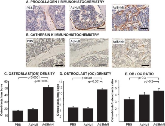
FIG. 3.

Osteoblasts and osteoclasts increase in vertebrae 18 days after AdShhN treatment. (A) Immunohistochemical staining for procollagen I, a cytoplasmic marker of osteoblasts: PBS (left); AdNull (middle); and AdShhN (right). Brown indicates positive staining. Bar = 40 μm. (B) Immunohistochemical staining for cathepin K, a marker of active osteoclasts: PBS (left); AdNull (middle); and AdShhN (right). Bar = 40 μm. (C–E) Quantification of osteoblast and osteoclast numbers (n = 5 for all groups). (C) Osteoblasts/mm of trabecular area. (D) Osteoclasts/mm of trabecular area. (E) Osteoblast/osteoclast ratio. For C–E, data shown as mean ± SE.
