
FIG. 2.
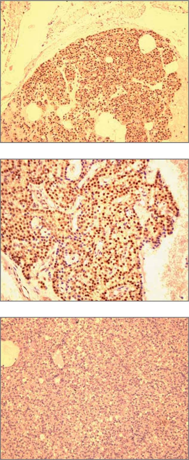
Immunohistochemical analysis of parafibromin expression. (Top) Normal parathyroid. A diffuse nuclear staining is present in most parathyroid cells (×200). (Middle) Parathyroid adenoma. The majority of cells show a positive nuclear staining (×400). (Bottom) Parathyroid carcinoma. Tumor cells show no nuclear staining (×200). (Reproduced from Eur J Endocrinol 156:547–554 with permission from the European Society of Endocrinology.)