
FIG. 5.
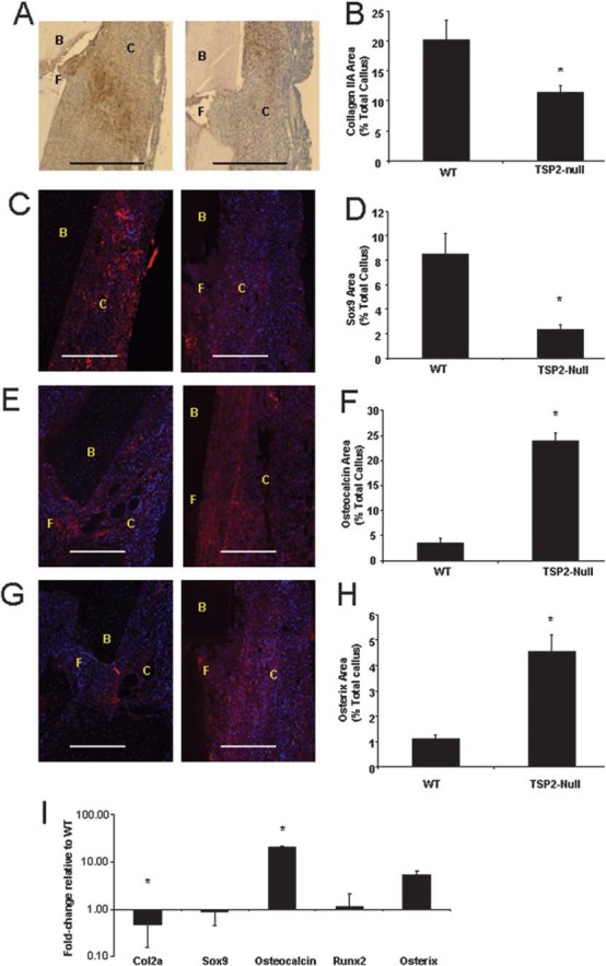
The fracture callus of TSP2-null mice 5 days after fracture has reduced chondrogenesis and increased osteoblast differentiation. Immunohistochemistry and histomorphometry were used to examine the differentiation of mesenchymal cells in the 5d calluses. (A and B) Type IIa collagen and (C and D) Sox9 expression were evaluated to determine areas of neochondrogenesis, whereas (E and F) osteocalcin and (G and H) osterix expression were used to indicate areas of new bone formation (magnification bar = 100 μm). Positive staining is either brown (A) or red (C, E, and G). Blue staining in C, E, and G represents DAPI-stained nuclei. B, bone; C, callus tissue; F, fracture. Values are mean ± SE of WT (n = 13) and TSP2-null (n = 12) mice. *Significantly different from WT; p < 0.05. (I) RNA was extracted from day 5 calluses and gene expression was evaluated using quantitative real-time PCR. Values are mean ± SE of fold-change in TSP2-null (n = 6) compared with WT mice (n = 5); *ΔCT significantly different from WT (p < 0.05).