
FIG. 6.
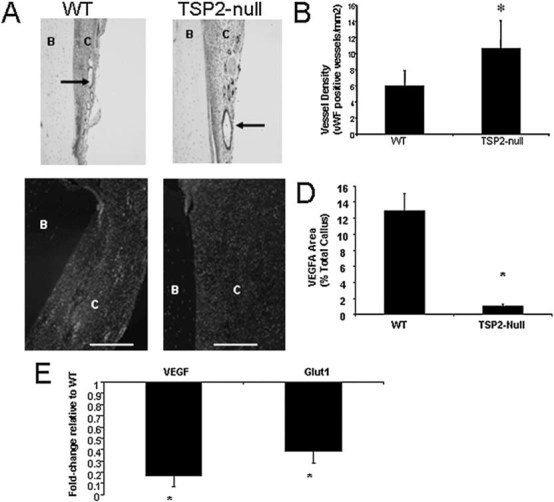
TSP2-null fractures show increased angiogenesis and a reduction in markers associated with HIF1-α activity. (A and B) Blood vessel density was determined by counting the total number of vWF-positive vessels in the callus (arrows). (B) Blood vessel density is 75% greater in the callus of TSP2-null mice compared with WT mice. (C and D) VEGF expression in calluses was evaluated using immunofluorescence (magnification bar = 100 μm). B, bone; C, callus tissue; F, fracture. Values are mean ± SE of WT (n = 13) and TSP2-null (n = 12) mice. *Significantly different from WT (p < 0.05). (E) RNA was extracted from day 5 calluses and gene expression was evaluated using quantitative real-time PCR. Values are mean ± SE of fold-change in TSP2-null (n = 6) compared with WT mice (n = 5). *ΔCT significantly different from WT (p < 0.05).