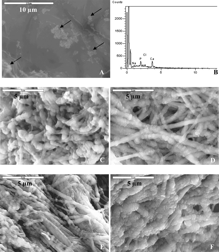
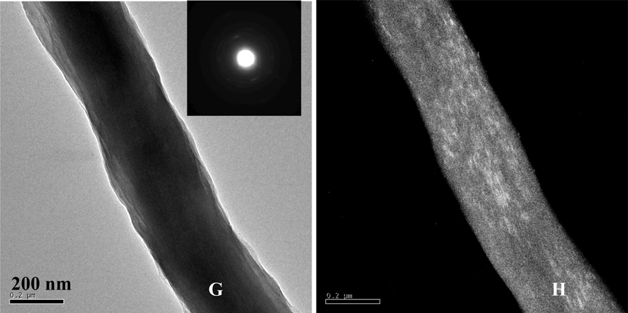
Figure 6.
High-magnification SEM of PILP-mineralized collagen samples using poly-L-glutamic acid (30-kDa), and combinations of PASP and PGLU as the process-directing agents. SEM (A) and EDS (B) of the surface of mineralized collagen sponge treated with PILP solution containing 50 µg/mL of PGLU30 for 6 days. Spherulitic clusters of hydroxyapatite are observed on the surface of collagen sponges treated with PGLU30 (arrows). (C–F) Comparison of low versus high molecular weight combinations. (C) SEM showing the inner-section of mineralized collagen sponge treated with PILP solution containing 50 µg/ml PASP14 and 50 µg/mL of PGLU15 for 3 days, and (D) for 6 days; (E) SEM of the inner-section of sample treated with PILP solution containing 50 µg/mL of PASP27 and 50 µg/mL of PGLU30 for 3 days, and (F) for 6 days. (G) Bright- and (H) dark-field TEM micrographs of a PILP-mineralized collagen fibril using the PASP27/PGLU30 combination. Inset: SAED pattern of mineralized fibril shown in (G).


