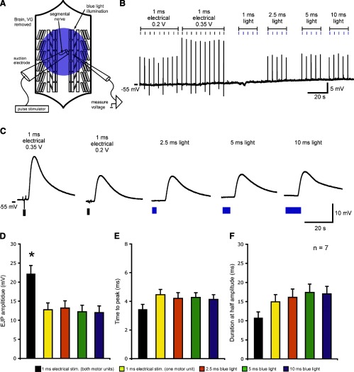
Fig. 3.
Comparison of light and electrically evoked excitatory junctional potential (LEJPs and EEJPs, respectively) in the absence of motor neuron cell bodies and ventral ganglion (VG) circuitry. A: schematic of a dissected larval preparation. The brain and ventral ganglion were removed. A single segmental nerve was stimulated via suction electrode. Larval muscle 6 was targeted for recording. B: long time-base recording showing a typical experiment. One motor unit was recruited with the lowest stimulus voltage. An additional motor unit was recruited as the electrical stimulus intensity increased. LEJPs were evoked by 2.5- to 10-ms light pulses. C: expanded time-base views of EEJPs and LEJPs shown in B. D–F: LEJPs showed amplitudes and time courses that were not statistically different from EEJPs evoked by the low-threshold motor unit (F > 0.05 by one-way ANOVA). Data from 1-ms light pulses are not shown because they did not evoke LEJPs in any preparations. In pooled data, resting membrane potentials were between −40 and −55 mV. Resting membrane potentials were not significantly different across stimulation types (F > 0.05 by one-way ANOVA; data not shown). Pooled data are presented as means ± SE. *Significant difference compared with all other conditions (P < 0.05 by one-way ANOVA with the Tukey-Kramer post hoc test).

