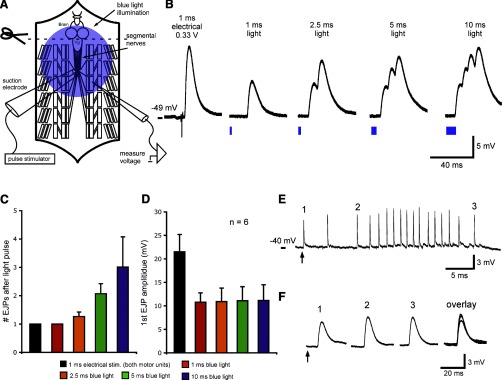
Fig. 4.
Comparison of LEJPs and EEJPs with motor neuron cell bodies and ventral ganglion intact. A: schematic of a dissected larval preparation, showing the brain, ventral ganglion segmental nerves, and an intracellular electrode in larval muscle 6. The brain is removed, but the ventral ganglion is intact. B: EJPs in response to a 1-ms electrical stimulus and four different blue light pulse durations. The electrical stimulus intensity was adjusted to activate both motor units innervating larval muscle 6. Note the multiple summating LEJPs after longer light pulse durations. C: number of EJPs for each light pulse duration. D: increasing light pulse durations did not affect the amplitudes of leading LEJPs. E: short light pulses can trigger long trains of spontaneous EJPs. A 1-ms light pulse (arrow) triggered a single EJP in larval muscle 6 (1) followed by a train of endogenously generated EJPs (2 and 3). F: LEJP was similar in amplitude and duration to spontaneous EJPs. Data in B, E, and F are from two different animals. In pooled data, resting membrane potentials were between −40 and −55 mV. Leading EJP amplitudes and resting membrane potentials were not significantly different across stimulation types (F > 0.05 by one-way ANOVA). Pooled data are presented as means ± SE.

