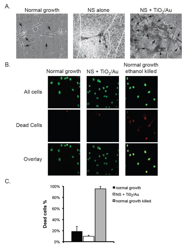
Figure 2.
Growth of normal human osteoblasts on nanosprings. A. Osteoblasts were grown for 3 days on plain glass (normal growth), NS or on NS coated with titania (TiO2) and gold nanoparticles (Au) and imaged by contrast light microscopy. Arrows point at individual cells. B. Cells grown on normal growth conditions or on NS + TiO2/Au surfaces for 5 days were stained with Vybrant Green (green), which labels the nuclei of all cells and with propidium iodide (red), which preferentially stains dead cells. The overlap of both stains is shown in the last row. As a positive control for death, cells grown on normal conditions were treated with ethanol (bottom row). C. The percentage of cells dead cells for the three types of cultures in B was determined for a total of 500 cells by dividing the number of propidium iodide positive cells by the number of Vybrant Green positive cells. Data were compiled from 3 independent experiments. Error bars represent standard deviation.

