Figure 4.
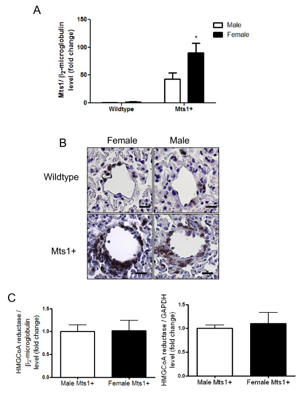
Mts1 expression is increased female Mts1+ mice compared to males. A: Mts1 gene expression is increased in the lungs of Mts1+ mice as assessed by qRT-PCR. This is exaggerated in lungs from female Mts1+ mice compared to male Mts1+ mice (n = 8). *P < 0.05 cf male Mts1+ mice B: Representative images of pulmonary arteries (x400 magnification) from female wildtype mice, male wildtype mice, female Mts1+ mice and male Mts1+ mice stained for Mts1. Mts1 expression is localised to the medial and adventitial layers of non-plexiform like pulmonary arteries and is most pronounced in female Mts1+ mice. Scale bars represent 20 μm. C: qRT-PCR analysis shows expression of the Mts1 promoter HMGCoA reductase is not influenced by gender in Mts1+ mice. Both β2-microglobulin and GAPDH were used as endogenous controls.

