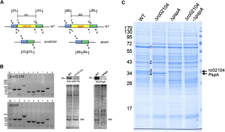
Fig. 4.
Gene deletion demonstrates that ro02104 and PspA are two of the major proteins of lipid droplets. A: The diagrams show in-frame deletions of ro02104 and pspA. The detailed construction and screening process were described (see supplementary Fig. II). B: The left panel shows PCR confirmation of the deletion mutants. 1–3, Target gene, including the upstream and downstream fragments, amplified using the mutagenic plasmid, or genomic DNA of the deletion mutant or WT as the template and primers a and d; 4–5, the region upstream of the target gene amplified using genomic DNA of the WT or the deletion mutant as the template and primers a and b; 6–7, the downstream region of the target gene amplified using genomic DNA of the WT or deletion mutant as the template and primers c and d; 8–9, the target gene from the WT and the deletion mutant amplified using genomic DNA of the WT or the deletion mutant as the template and primers f and r. The right two panels show the Western blotting results that verify the ro02104 and pspA deletion mutants. C: LD proteins were extracted from the WT, ro02104 mutant, pspA mutant, and ro02104-pspA double-deletion mutant, and separated by 10% SDS-PAGE followed by Colloidal Blue staining. The main LD protein bands are labeled 1, 2, 3 (ro02104), and 4 (PspA).

