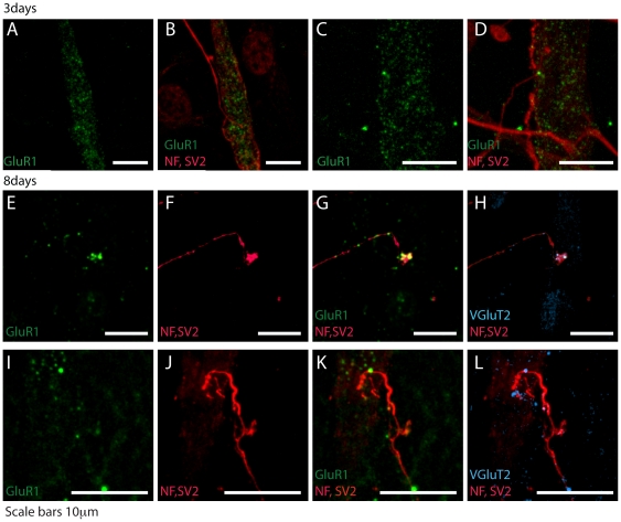
Figure 3. Time course of synaptic contacts formation in cocultures.
Examples of cortical neurons cocultured with myotubes for 3 (A–D) and 8 days (E–L), fixed, immunostained and studied by confocal microscopy. AMPARs (GluR1 subunit) are in green, axonal neurofilaments and terminations are in red (NF, SV2). At 3 days AMPARs are diffusely distributed (A, C) and myotubes often receive multiple synaptic contacts (B, D). At 8 days AMPARs form clusters (E, I) near or under the terminations (G, K). The Vesicular Glutamate transporter 2 (VGluT2, blue) confirm that the synaptic contact is glutamatergic (H, L). Scale bars 10 µm.

