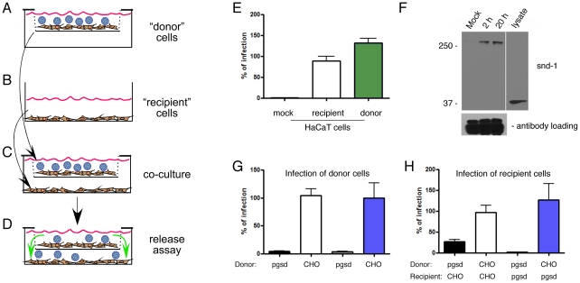
Figure 4. Released HMW complexes including HSPG and HPV16 are required for infection.
(A–D) Schematic of the “donor” cell/“recipient” cell co-culture system indicating how cells were exposed to PsVs. PsVs were allowed to bind donor cells without internalization (A). Donor cells on coverslips were washed thoroughly to remove unbound PsVs and transferred to mesh inserts above the recipient cells (B) to co-culture with gentle rocking for 24 h and allow released HPV complexes from donors to access the recipient cells (C–D). All experiments employed CM. (E) Relative HPV16 infection levels of HaCaT donor cells and recipient HaCaT cells compared to mock infected cells as verification of the co-culture virus release model. (F) Non-reducing SDS-PAGE and immunoblot of syndecan-1 (polyclonal rabbit antisera) following IP of HPV16 (mouse monoclonal anti-L1). IP was performed by immobilizing anti-L1 in the lower chamber in place of cells (see panel B) to capture HPV16 released from mock exposed cells (M) or HPV16-exposed HaCaT donor cells at 2 or 20 h post virus exposure. Lower panel IgG detection is included as a loading control. (G) Relative infection levels in CHO-K1 and pgsd-677 cells used as donor cells bound to HPV16 PsV and co-cultured above the recipient cells. (H) Relative infection levels in CHO-K1 and pgsd-677 cells used as recipient cells co-cultured below the PsV-bound donor cells corresponding to the data in panel G. Infectivity data (E,G,H) were normalized to the mean value of the infected control set to 100% and represent the mean ± SEM of 4 replicate infections.

