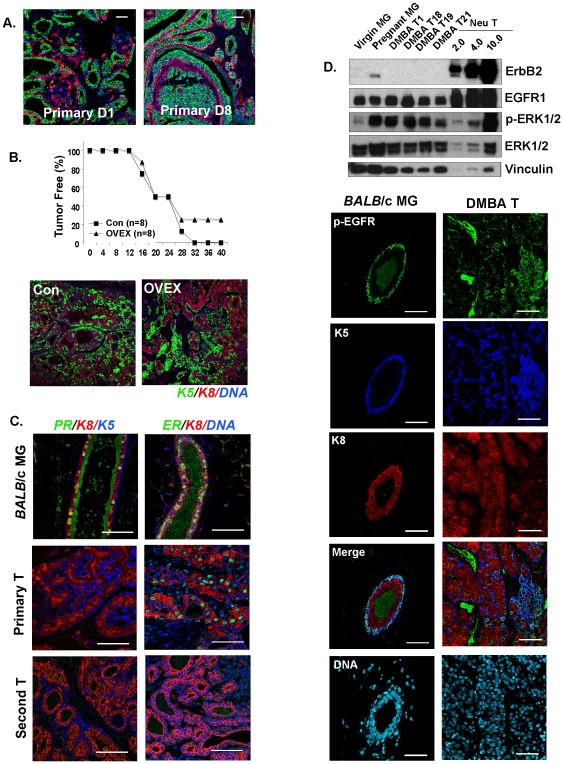
Figure 1. KRT5+ DTumors parallel human basal-like breast tumors by functional and molecular assay.
(A) Mixed lineage tumors. 19 primary tumors were double stained with lineage-specific markers (keratin 5, basal cells, stained red; keratin 8, luminal cells, stained green) and 17 primary tumors (90%) comprise ≥10% K5-positive cells, as well as K8-positive cells, indicating that these tumors are bi-lineal. Two representative images from two different primary tumors are shown. Scale bar = 50 µm. (B) Estrogen-independence. The appearance of palpable tumor masses was measured after transplanting 10,000 tumor cells into either control or ovarectomized three-week old BALB/c recipients (representative of 3 strains of primary tumor). Re-growth of tumors is no different in recipients grafted at 3 weeks of age. Paraffin sections from tumors removed from ovariectomized hosts were compared to tumors from normal hosts, by double staining for K5 (green) and K8 (red). There was no difference in their basal/luminal constituent cell types. (C) Experimental tumors are ERα/PR-negative. Paraffin sections from normal virgin BALB/c mammary glands (MG), together with an example of a primary tumor (Primary T) and a secondary tumor (Second T), were stained for ERα and PR, and counterstained as indicated with either K5 (basal) or a DAPI nuclear stain. (The specificity of the anti-PR-A staining procedure is illustrated; Fig. S7). (D) EGF signaling receptor expression. To evaluate expression of erbB2 and EFGR1, together with a downstream effector of EGFR1, p-ERK1/2 (and total ERK1/2) in four primary tumors (DMBA D1, D18, D19, and D21), tumor tissue lysates (20 µgs) were compared with tumor tissue lysates from erbB2/neu transgenic mice (with 10.0, 4.0 and 2.0 µgs total protein), and with mammary gland from mid-pregnant and virgin mice. Vinculin was used as a loading control. Immunohistochemical staining for pEGFR in paraffin sections from normal mammary glands and tumors confirmed and extended the Western blotting results. Cell surface-associated pEGFR is typical of basal cells in normal mammary glands and this cell type-specific expression pattern is conserved in basaloid tumor cells. (Note that the green stain in the lumens is an artifact associated with sticky luminal secretions; panels C and D).

