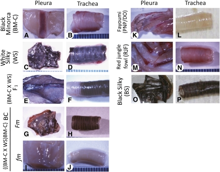
Figure 1 .
Pigmentation of pleura and trachea in several chicken lines. WS and BS have heavily pigmented internal organs whose colors are clearly different from those in the fm lines (BM-C, PNP/DO, and RJF). F1 between BM-C and WS shows Fm, and BC progeny between the F1 and BM-C are classified into Fm or fm groups. (A and B) BM-C. (C and D) WS. (E and F) F1. (G and H) BC judged as Fm. (I and J) BC judged as fm. (K and L) PNP/DO. (M and N) RJF. (O and P) BS. (A, C, E, G, I, K, M, and O) Pleura. (B, D, F, H, J, L, N, and P) Trachea. (Size) one pitch of the scales: 1 mm.

