
Figure 3.
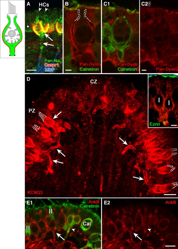
Domain 3, comprising the basolateral calyx and the stretch of axon leading to the heminode, selectively expressed NaV channels, KCNQ3, and certain scaffolding proteins. A, Pan-NaV antibody produced a complex pattern of label, including domains 1 and 3 (arrows) and the heminode (domain 4), as well as type I hair cells (arrowheads) and adjacent type II hair cells (asterisk). B, C, Pan-dystrophin (Pan-Dystr, red) labeled domain 3 of dimorphic (calretinin-negative), but not calyx-only (calretinin-positive, green) afferents (compare B with C1 and C2). D, Domain 3 in peripheral-zone dimorphic afferents was intensely immunoreactive for KCNQ3, as shown by this flattened projection of a stack of confocal images. KCNQ3 staining (arrows) extended throughout domain 3 from the basolateral outer surface of the calyx to the heminode. Note the lack of stain in domain 2, marked by dashed white lines. The inset shows a higher magnification single image of a dimorphic afferent with a “complex” calyx ending that envelops two adjacent type I hair cells. Antibody to ezrin (green) labeled the apical microvilli of the sensory epithelium and the heminode, where it combined with the red KCNQ3 immunolabel to form a yellow band. E, AnkyrinB antibody labeled domain 3 in dimorphic afferents (E1, E2, red, long arrows) but not calyx-only afferents (E1, E2, green, arrowheads). Scale bars: A, D, E, 10 μm; D, inset, 5 μm; B, C, 2 μm.