
Figure 1.
Mutations in RHBDF2 Underlie Tylosis
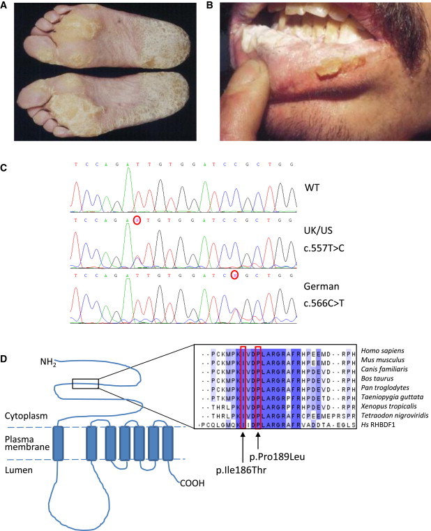
Clinical images of a tylosis patient showing the focal nonepidermolytic palmoplantar keratoderma (A) and oral leukokeratosis (B). Sanger sequence traces (C) displaying the c.557T>C mutation identified in the UK and USA families and the c.566C>T mutation identified in the German family.
(D) Schematic illustrating of the structure of RHBDF2, a seven-transmembrane-domain protein, and the approximate location of the alterations identified in tylosis patients. Protein alignment with ClustalW illustrates that the amino acid residues mutated in the tylosis patients (in p.Ile186Thr and p.Pro189Leu) are highly conserved across a wide range of eukaryotic species as well as in RHBDF1, a closely related member of the iRhom family.