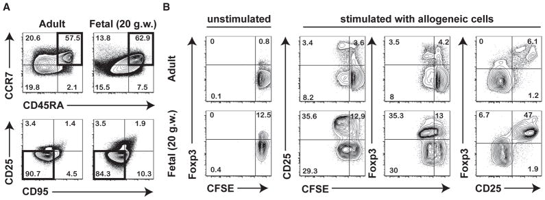
Fig. 1.
Fetal naïve CD4+ T cells display functional differences compared to adult naïve CD4+ T cells. (A) Flow cytometric analysis of the phenotype of naïve CD4+ T cells isolated from fetal mLNs (18–22 g.w.) and adult peripheral blood mononuclear cells (PBMC) (25–35 y.o.). Panels labeled (i) depict initial gating on CD3+CD4+ T cells showing CD45RA vs. CCR7 staining and those labeled (ii) depict CD45RA+CCR7+ cells (highlighted in black in panel i) subsequently gated on CD25 CD95− cells that are considered “naïve” CD4+ T cells (highlighted in black in panel ii). Data are representative of at least 3 independent donors. (B) Sorted naïve CD4+ T cells were either left unstimulated, or cultured for 6 days in the presence of irradiated allogeneic PBMC. Proliferation of fetal and adult naïve CD4+ T cells was measured by CFSE dilution and analyzed by flow cytometry. Tregs were phenotypically identified by upregulation of CD25 and Foxp3.

