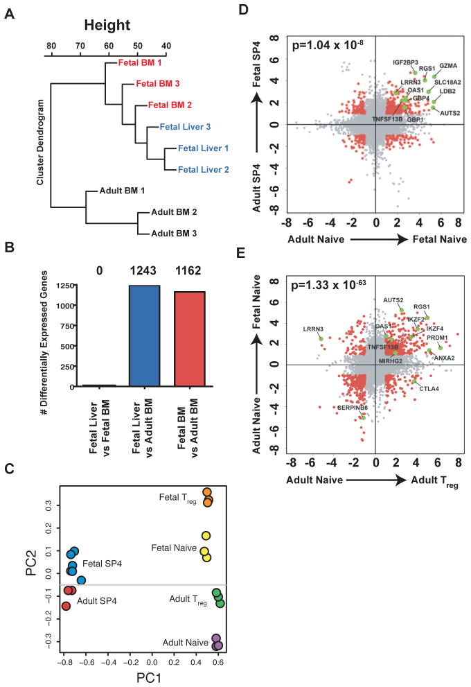
Fig. 4.
Fetal and adult HSPC give rise to SP4 thymocytes with distinct gene signatures. (A) Unbiased cluster analysis based on gene expression for SP4 thymocytes from fetal BM, fetal liver, and adult BM hematopoietic progenitors.(B) Total number of genes found to be significantly different comparing SP4 thymocytes derived from fetal and adult progenitor populations. (C) Principle component analysis showing clear separations between SP4 thymocyte populations and peripheral T cells (PC1) as well as differences between fetal and adult SP4 and peripheral naïve CD4+ T cell populations (PC2). (D) Scatter plot depicting genes that are differentially expressed between fetal and adult SP4 thymocytes, and between fetal and adult peripheral naïve CD4+ T cells. (E) Scatter plot depicting genes that are differentially expressed between adult and fetal naïve T cells, and between adult naïve and adult Tregs. For both (D) and (E), differential expression was determined using a false discovery rate of ≤ 5% and fold-change of ≥ two-fold. The significance of overlap between groups of genes was determined using a chi-squared test for independence.

