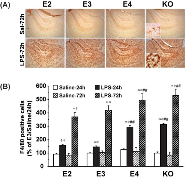
Figure 2. LPS induces F4/80 positive cell activation in an APOE genotype dependent manner in the hippocampus.

APOE2, APOE3, APOE4 and APOE knock-out mice were sacrificed 24h and 72h after ICV injection of LPS (1000ng) or saline. (A) Representative IHC images of F4/80-positive microglia in the hippocampus under 5X and 63X lens (Inset). The scale bar is 100μm and 10μm, respectively. (B) Quantification of F4/80- positive cells in the hippocampus. The numbers of F4/80-positive microglia are expressed as mean ± SD (n=4-5/group). **P<0.01, compared with saline treatment at the same time point. ##P<0.01, compared with APOE2 or APOE3 at the same LPS treatment time point.
