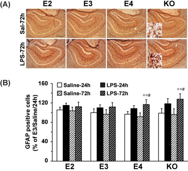
Figure 4. LPS effect on hippocampal astrocyte activation.

APOE2, APOE3, APOE4 and APOE knock-out were sacrificed 24h and 72h after ICV injection of LPS (1000ng) or saline. (A) Representative IHC images of GFAP-positive astrocyte in the hippocampus under 5X and 63X lens (Inset). The scale bar is 100 μm and 10μm, respectively. (B) Quantification of GFAP-positive astrocyte numbers in the hippocampus. Data are expressed as mean ± SD (n=4-5/group). **P<0.01, compared with saline treatment at the same time point. #P<0.05, compared with APOE2 or APOE3 with LPS treatment at 72 hours.
