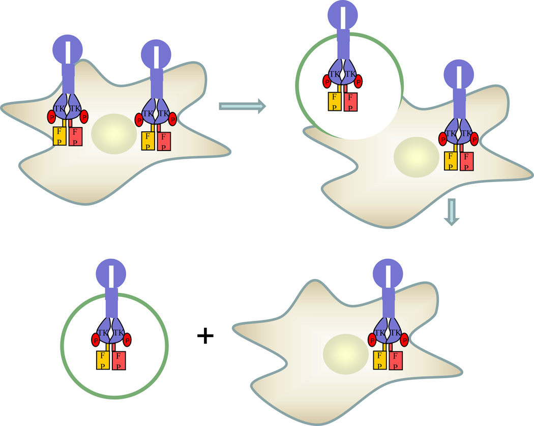
Figure 3.
Overview of RTK dimerization measurements in plasma membrane-derived vesicles. Cells are transfected with genes encoding RTKs fused to fluorescent proteins, and then vesiculated using established protocols. The vesicles are imaged in a confocal microscope, acquiring donor, acceptor and FRET images for each vesicle. The QI-FRET method, described in (53–55), is used to determine the donor and acceptor concentration, and the FRET efficiency in each vesicle. This information is then used to determine the concentrations of monomers and dimers in each vesicle, the dimerization constant K1 and the free energy of dimerization.

