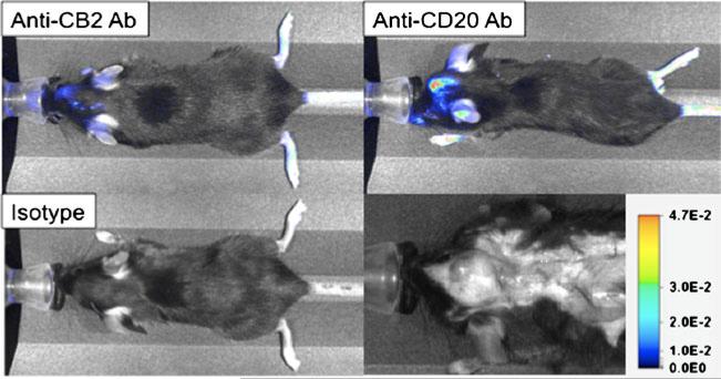
Fig. 4.
Lymphocytes and activated leukocytes can be tracked and imaged non-invasively using NIRF imaging in mouse models of brain tumors. Three antibodies (anti-cannabinoid receptor 2, anti-CD20 and IgG2a isotype control) were labeled with LiCOR IRDye800-NHS and purified using Sephadex G-25 desalting resin. Female C57bl6 bearing intracranial 3LL Lewis Lung carcinoma cells (day 6 post-implantation) were then injected intra-peritoneally with 10 mg of optically-labeled antibody as indicated in PBS. At 72 h post-antibody injection, each mouse was then imaged using a LiCOR Pearl Impulse imager exciting at 745 nm and detecting at 800 nm. Images show accumulation of anti-CB2 Ab at the site of the 3LL brain tumor (tumor in red arrow in bottom left white-light photo) showing the presence of activated macrophages and microglia in the vicinity of the tumor. Similarly, strong accumulation of anti-CD20 Ab indicates the presence of numerous B lymphocytes present in or around the tumor. Negative isotype control antibody presence within the tumor (but present in spleen, data not shown) indicates specific binding by anti-CB2 and anti-CD20 antibodies

